

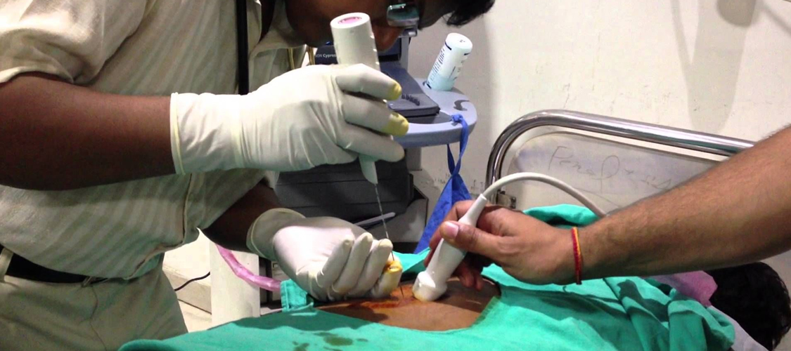
Biopsy is a method of inserting a special needle into a tumour tissue, and get samples; this is done under local anesthesia to ensure painless experience for the patient using CT or ultrasound guidance; the continuous monitoring of the needle during the procedure ensures that the needle reaches the correct spot.
FNAC is method of inserting tiny needle into small lesions, eg: thyroid nodule; this is done under ultrasound guidance to ensure that the needle reaches the area of high cellularity avoiding necrotic and fluid areas. The slides are made from the needle aspirate and sent for cytologist interpretation.
Drainage: Pus collection in the body can lead to continuing illness, the pus has to be let out and in the past, this involved complex surgeries, with advent of CT and ultrasound, a special needle can be inserted into these collections (“abscess”) and pus can be aspirated without disturbing other structures. Further microbiology tests on the pus, give the treating doctor information about the “bacteria / bug” and the appropriate “Antiobiotic” that helps to cure the bug.
This team has done over 5000 biopsies / FNAC / Abscess drainage with successful results.
Needle biopsy
During a needle biopsy, your doctor uses a special needle to extract cells from a suspicious area.
A needle biopsy is often used on tumors that your doctor can feel through your skin, such as suspicious breast lumps and enlarged lymph nodes. When combined with an imaging procedure, such as X-ray, needle biopsy can be used to collect cells from a suspicious area that can’t be felt through the skin.
Needle biopsy procedures include:
- Fine-needle aspiration. During fine-needle aspiration, a long, thin needle is inserted into the suspicious area. A syringe is used to draw out fluid and cells for analysis.
- Core needle biopsy. A larger needle with a cutting tip is used during core needle biopsy to draw a column of tissue out of a suspicious area.
- Vacuum-assisted biopsy. During vacuum-assisted biopsy, a suction device increases the amount of fluid and cells that is extracted through the needle. This can reduce the number of times the needle must be inserted to collect an adequate sample.
- Image-guided biopsy. Image-guided biopsy combines an imaging procedure — such as X-ray, computerized tomography (CT), magnetic resonance imaging (MRI) or ultrasound — with a needle biopsy.Image-guided biopsy allows your doctor to access suspicious areas that can’t be felt through the skin, such as abnormalities on the liver, lung or prostate. Using real-time images, your doctor can make sure the needle reaches the correct spot.
You’ll receive a local anesthetic to numb the area being biopsied in order to minimize the pain.