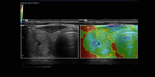

Elastography helps visualize the difference between the biomechanical properties of normal and diseased tissues. In that sense, Elastography is similar to palpation. To be palpable, the object must be harder or softer than the tissue surrounding it. Fingers can displace tissue downward during palpation, and the pressure receptors on the skin of the fingers can sense the local stress values. Stress is higher on the fingers overlying a superficial hard lesion and lower on the fingers overlying softer tissues.
The same principle can be used with ultrasound, with the transducer used in the same manner as a finger. Hard lesions will move as a unit, and soft lesions will deform under the local pressure. We could use the ultrasound transducer for compression and measure the relative flattening of the lesion compared with the thinning of an adjacent tissue layer.
In 1991, ultrasound Elastography was introduced that involved a small external compression and measured the tissue response with the use of cross-correlation methods. Orphic called this method Elastography . This method of imaging tissue hardness is based on the Young elastic modulus. Pre- and post compression ultrasound images of the same tissue are recorded, and tissue movement during compression is analyzed to estimate strain .
The basic principle of Elastography can be summarized as follows. First, disturb the tissue with a physical source. Second, measure the resulting mechanical response and infer the bio-mechanical properties of the underlying tissue by applying mechanical models to the measured mechanical response. Disturbing the tissue with a physical source can be achieved by quasi static, harmonic, or transient sources. Quasi static Elastography uses external compression as the strain source and then visualizes the strain induced in tissue with the use of either an external or internal source. In harmonic Elastography , low-frequency acoustic waves are transmitted within the tissue. The phase and amplitude of the propagating waves are visualized using color Doppler imaging . In transient Elastography , an ultrasound scanner with a high frame rate (i.e., 10,000 frames per second) is used to generate and then track the propagation of shear waves in tissue . Shear waves are tissue movements perpendicular to the direction of the ultrasound wave transmission.
Ultrasound Elastograph is one of the latest technology that allows the sound to ‘palpate’ the lesion.
Applications
- Early detection of liver fibrosis – in fatty infiltration of liver.
- Additional tool for detection of breast cancer, thyroid cancer and lymph node metastasis